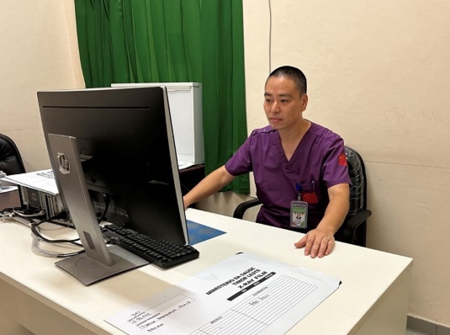
Here, we work alongside local healthcare workers and medical professionals from other countries, learning from each other and providing high-quality medical services to the people of Timor-Leste.
The healthcare system in Timor-Leste is generally underdeveloped. Although there are some aspects we, as Chinese doctors, can learn from, there are still many areas that need improvement. In light of this, the Chinese Medical Team hopes to create a series of health forums, where doctors from various specialties will give talks, exchange experiences with colleagues in Timor-Leste, and provide health education to the people of Timor-Leste. I am Dr. Luo Wei, the leader of the Chinese Team. I come from the First Affiliated Hospital of Chengdu Medical College, and my specialty is medical imaging. Today, I will start by giving you a talk on the topic of medical imaging examinations.
Medical imaging technology plays a key role in modern healthcare. Different imaging methods can help doctors accurately diagnose diseases. However, due to the unique characteristics of each imaging modality, selecting the appropriate method is crucial, especially in resource-limited environments like Timor-Leste.
X-ray
(a) Indications:
- Bone diseases: fractures, dislocations, arthritis.
- Chest diseases: pneumonia, tuberculosis, pneumothorax.
- Dental examinations: used to check cavities and root problems.
(b) Advantages:
- Economical: low cost, simple equipment, easily accessible in primary care hospitals.
- Quick diagnosis: fast examination, especially for the preliminary diagnosis of acute fractures and lung infections.
(c) Disadvantages:
- Radiation risk: although the radiation dose is small, frequent use should be avoided, particularly in children and pregnant women.
- Poor soft tissue imaging: X-ray is less effective in displaying soft tissues such as muscles and internal organs.
Ultrasound
(a) Indications:
- Abdominal examinations: liver, gallbladder, kidneys, spleen, etc.
- Obstetric and gynecological exams: fetal development, uterine and ovarian diseases.
- Cardiac and vascular examinations: structural abnormalities in the heart, blood vessel narrowing, etc.
(b) Advantages:
- No radiation: ultrasound uses sound waves to create images, making it safe for pregnant women and children.
- Real-time dynamic imaging: suitable for evaluating fetal heartbeats and dynamic organ changes.
- Portable equipment: easy to promote in primary care hospitals, with relatively low cost.
(c) Disadvantages:
- Limited penetration: unable to penetrate gas or bones, making it unsuitable for lung or bone disease assessments.
- Image quality depends on the operator: the results of an ultrasound exam largely depend on the operator's skill and experience.
Computed Tomography (CT)
(a) Indications:
- Brain examinations: intracranial hemorrhage, brain tumors, stroke.
- Thoracoabdominal diseases: lung nodules, liver tumors, acute abdominal conditions.
- Complex fractures: used to assess multiple fractures or joint structures.
(b) Advantages:
- High-resolution imaging: provides detailed cross-sectional images, suitable for evaluating complex conditions.
- Fast imaging: particularly useful for emergency patients, such as those with intracranial hemorrhage or traumatic fractures.
(c) Disadvantages:
- High radiation: compared to X-rays, CT involves a higher radiation dose, so it should be used with caution.
- High cost: CT equipment is expensive, and the examination cost is higher, making it less suitable for resource-limited primary care hospitals.
Magnetic Resonance Imaging (MRI)
(a) Indications:
- Central nervous system diseases: brain tumors, spinal cord injuries.
- Joint and soft tissue injuries: ligament tears, cartilage damage.
- Cardiac and vascular diseases: cardiomyopathy, aneurysms.
(b) Advantages:
- No radiation: MRI does not use ionizing radiation, making it safer.
- Excellent soft tissue imaging: MRI can clearly show the brain, spinal cord, muscles, and joints, making it ideal for diagnosing complex conditions.
- Multi-plane imaging: MRI can provide high-resolution images from multiple angles.
(c) Disadvantages:
- Long examination time: compared to X-rays and CT, MRI takes longer.
- Sensitivity to metal: patients with metal implants (such as pacemakers) cannot undergo MRI examinations.
- High cost: MRI equipment and examination costs are high, making it difficult to promote in resource-limited settings.
Conclusion
In the healthcare situation of Timor-Leste, choosing the appropriate imaging method depends on the patient's condition, the availability of equipment, and the specific needs of the examination. X-ray and ultrasound are the most commonly used methods in primary care institutions, due to their low cost and widespread accessibility. CT and MRI are more suitable for complex cases, offering more detailed images, but they should be used judiciously due to their higher cost. By making reasonable use of these diagnostic tools, we can provide patients with more accurate diagnoses and improve the quality of medical services.


